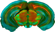
We address these questions at the cellular and circuit level, by performing long-term calcium imaging experiments in the primary and higher visual cortical areas and in the medial prefrontal cortex of the mouse. We make use of two-photon microscopy to image calcium activity of large groups of neurons throughout the process of learning visual categories. By quantifying changes in function of neurons within specific brain areas, we aim to determine different stages of the process of forming abstract/complex representations.
Conventional two-photon microscopes are large and heavy setups and cannot easily be adopted to behavioral paradigms that require the mouse to roam-around freely, such as place-reward learning. Therefore, we additionally make use of a head-mounted miniaturized epifluorescence microscope that the mouse can carry around while performing maze-learning tasks.
Using these approaches, we identify specific cells and circuits in the brain that are involved in learning of episodic or semantic associations. In subsequent steps, we aim to trace connectivity within, and the inputs to, these circuits in order to better understand how learned representations arise from changes in the underlying synaptic connectivity.


