Golgi-staining of pyramidal cells in an organotypic culture of rat cortex (pseudo-phasecontrast microscopy). ©Tobias Bonhoeffer
 Magnification of the layer staining shown on the left, in the original colors: calbindin in green, Ctip2 in red, and cell nuclei in blue. ©Volker Staiger, Simon Weiler
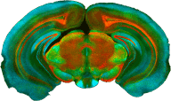
 Immunostaining for calbindin and Ctip2 to distinguish between the different layers in a coronal section of a mouse brain. ©Volker Staiger, edited by Julia Kuhl
 Pyramidal cells in hippocampal region CA1 of a GFPm mouse. Cell nuclei are stained using DAPI (blue), and the cell bodies using Nissl staining (red). ©Claudia Huber
 Section of mouse cortex stained with the Golgi-technique to show pyramidal cells. ©Volker Staiger
 Pseudo-phasecontrast microscopy of mouse hippocampus. The pyramidal cells are stained with the Golgi technique. ©Volker Staiger
 Golgi-staining of pyramidal cells in the cortex of a coronal section of a mouse brain. ©Tobias Bonhoeffer
 Pyramidal cells (green) in layer 5 of the cortex in a GFPm mouse. The Nissl staining (red) shows cell bodies. ©Volker Staiger, Robert Kasper
 3D-Print of a dendrite with spines reconstructed from an electron microscopical image stack. ©Volker Staiger, Max Sperling
 Hippocampal neurons in organotypic slice cultures stained with dsRed using the Gene-Gun technique. Fluorescent intensity is color-coded. ©David Laubender
 Hippocampal neurons in organotypic slice cultures stained with dsRed using the gene-gun technique. Fluorescent intensity is color-coded. ©David Laubender
 Immuno-staining of hippocampal area CA1 in a coronal section of the mouse brain. Blue: cell nuclei stained with DAPI; green: GFAP immunostaining showing astrocytes; red: microglia cells stained with Iba1. ©Volker Staiger
 Dendrite with spines in the mouse cortex (Golgi-staining). ©Volker Staiger
 Immunostaining of an organotypic culture of rat hippocampus showing astrocytes (green, GFAP staining), microglia (red, Iba1) and nuclei (blue, DAPI). The purple spots are due to an overlay of blue and red. ©Volker Staiger
 Mouse hippocampal primary culture stained for drebrin (Antibody-staining, green) and for actin (phalloidin staining, red). Overlay of both in yellow. ©Volker Staiger
 Mouse hippocampal primary culture stained for microtubuli-associated-protein (MAP2, green) and drebrin (red). Cell nuclei are stained in blue. ©Volker Staiger